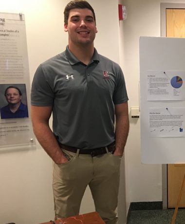
An engineering major works to develop a personalized cancer-detection system.
CELEBRATE! Week offers an annual opportunity to highlight the academic and artistic achievements of Elon students and faculty. Each day this week, we'll be putting the spotlight on a student scholar's research — what they are seeking to find out, and who they became interested in their project.
 Name: Matthew Foster
Area of study: Biomedical Engineering
Major(s): Engineering
Minor(s): Physics, Math
Faculty mentor: Richard Blackmon, assistant professor of engineering
Title of research: "Building an Optical Coherence Tomography Imaging System for Personalized Cancer Treatment"
Abstract:
Early detection and treatment remain our best tool against cancer. One potential method of detecting cancer uses Optical Coherence Tomography (OCT), a minimally invasive technology that uses near-infrared light to create high-resolution images in biological tissue.
My research works to develop a form of cancer detection and treatment that can be personalized to an individual. The OCT system is used to identify the cancerous cell in a tissue sample. Once this sample is identified, a therapeutic laser is used to heat and kill the cancerous cells. By using this targeting method to only eliminate the cancerous cells, the treatment is personalized to each individual.
In previous work, I have built an OCT imaging system capable of taking cross-sectional images of breast and skin tissue. I have also created a user-friendly interface and software to control our OCT imaging machine so that it can be used by people with a limited understanding of computer software. In this work, I have combined my software into a master user interface capable of controlling all aspects of the system at once.
In future work, I will demonstrate the efficacy of the system by taking live images of biological samples and processing them in real-time. These images’ will be used to further calibrate my current OCT imaging system.
In other words:
This is a personalized cancer-detection system. It is meant to be much less invasive than traditional detection and treatment systems, allowing for personalized treatment that meets each individual person needs. This semester, I have worked to get the system running smoothly, and tested the imaging capabilities.
Explanation of study:
This research is a continuation of the research I did last summer in Elon’s SURE program. From the beginning, I have built the system and wrote the code for the user interface. The purpose of an easy-to-use interface is so that in the future, if a doctor or biologist who is not technically inclined wants to use the system for tests, they will be able to easily. My research this semester involved refining the user interfaces and tuning the system. This semester we were able to take our first live test images with the system. Studying those images allowed us to continue to refine and focus the system further.
What made this research interesting to you? How did you get started?
This research was interesting to me because I believe that biomedical engineering is a way to truly make peoples lives better through your designs. I enjoyed the challenge of tackling a tough problem, and working with a device that would be used to treat cancer patients was something that interested me.
How has undergraduate research contributed to your experience at Elon?
Applying concepts used inside of the classroom to a real-world system was exciting and eye-opening to me. This experience and the opportunity to perform such high-level research has greatly contributed to my learning experience here at Elon.


